| dc.contributor | Vall d'Hebron Barcelona Hospital Campus |
| dc.contributor.author | Díaz-Mercedes, Sherley |
| dc.contributor.author | Javier Aguirre, Jose |
| dc.contributor.author | Machado, Isidro |
| dc.contributor.author | Rodrigo, Maria Teresa |
| dc.contributor.author | Landolfi, Stefania |
| dc.contributor.author | Archilla, Ivan |
| dc.contributor.author | Tarragona, Jordi |
| dc.date.accessioned | 2022-01-13T17:11:30Z |
| dc.date.available | 2022-01-13T17:11:30Z |
| dc.date.issued | 2021-03 |
| dc.identifier.citation | Archilla I, Díaz-Mercedes S, Aguirre JJ, Tarragona J, Machado I, Rodrigo MT, et al. Lymph Node Tumor Burden Correlates With Tumor Budding and Poorly Differentiated Clusters: A New Prognostic Factor in Colorectal Carcinoma? Clin Transl Gastroenterol. 2021 Mar;12(3):e00303. |
| dc.identifier.issn | 2155-384X |
| dc.identifier.uri | https://hdl.handle.net/11351/6791 |
| dc.description | Prognostic factor; Colorectal Carcinoma; Tumor Burden |
| dc.description.abstract | INTRODUCTION:
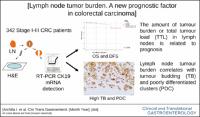
Molecular lymph node (LN) staging in early colorectal cancer (CRC) has demonstrated to be more precise than conventional histopathology pN staging. Tumor budding (TB) and poorly differentiated clusters (PDCs) are associated with LN metastases, recurrences, and lower survival in CRC. We evaluated the correlation between the total tumor load (TTL) in LNs from CRC surgical specimens with patient outcome, TB, and PDC.
METHODS:
In this retrospective multicentre study, 5,931 LNs from 342 stage I–III CRC were analyzed by both hematoxylin and eosin and molecular detection of tumor cytokeratin 19 mRNA by one-step nucleic acid amplification. TB and PDC were evaluated by hematoxylin and eosin and cytokeratin 19 immunohistochemistry.
Results:
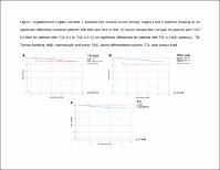
One-step nucleic acid was positive in 38.3% patients (n = 131). Tumor Budding was low in 45% cases, intermediate in 25%, and high in 30%. Poorly Differentiated Clusters were low-grade G1 in 53%, G2 in 32%, and G3 in 15%. TB and PDC correlated with TTL, high-grade, lymphovascular and perineural invasion, pT, pN and stage (P < 0.001). TB, PDC, and TTL ≥ 6,000 copies/µL were associated with worse overall survival (P = 0.002, P = 0.013, and P = 0.046) and disease-free survival (P < 0.001).
Discussion:
The implementation of more sensitive molecular methods to assess LN status is a promising alternative approach to pN staging, which could be integrated to other factors to help risk stratification and management of patients with early-stage CRC. This study demonstrates the correlation of the amount of LN tumor burden with TB and PDCs. TTL is related to the outcome and could be used as a new prognostic factor in CRC (see Visual Abstract, Supplementary Digital Content 2, https://links.lww.com/CTG/A512). |
| dc.language.iso | eng |
| dc.publisher | Lippincott Williams & Wilkins |
| dc.relation.ispartofseries | Clinical and Translational Gastroenterology;12(3) |
| dc.rights | Attribution-NonCommercial-NoDerivatives 4.0 International |
| dc.rights.uri | http://creativecommons.org/licenses/by-nc-nd/4.0/ |
| dc.source | Scientia |
| dc.subject | Còlon - Càncer - Prognosi |
| dc.subject | Recte - Càncer - Prognosi |
| dc.subject | Nodes limfàtics - Aspectes moleculars |
| dc.subject.mesh | Colorectal Neoplasms |
| dc.subject.mesh | /diagnosis |
| dc.subject.mesh | Lymph Nodes |
| dc.title | Lymph Node Tumor Burden Correlates With Tumor Budding and Poorly Differentiated Clusters: A New Prognostic Factor in Colorectal Carcinoma? |
| dc.type | info:eu-repo/semantics/article |
| dc.identifier.doi | 10.14309/ctg.0000000000000303 |
| dc.subject.decs | neoplasias colorrectales |
| dc.subject.decs | /diagnóstico |
| dc.subject.decs | ganglios linfáticos |
| dc.relation.publishversion | https://doi.org/10.14309/ctg.0000000000000303 |
| dc.type.version | info:eu-repo/semantics/publishedVersion |
| dc.audience | Professionals |
| dc.contributor.organismes | Institut Català de la Salut |
| dc.contributor.authoraffiliation | [Archilla I, Díaz-Mercedes S, Rodrigo MT] Pathology Department, Center of Biomedical Diagnosis (CDB), Hospital Clínic, University of Barcelona, IDIBAPS, Spain. [Aguirre JJ] Pathology Department, Arava University Hospital, Vitoria-Gasteiz, Spain. [Tarragona J] Pathology Department, Hospital Arnau de Vilanova, Lleida, Spain. [Machado I] Pathology Department, Instituto Valenciano de Oncologia and Hospital QuironSalud, Valencia, Spain. [Landolfi S] Department of Pathology, Hospital Universitario Torrecardenas, Almeria, Spain. Servei de Patologia, Vall d’Hebron Hospital Universitari, Barcelona, Spain |
| dc.identifier.pmid | 33939382 |
| dc.identifier.wos | 000681351500003 |
| dc.rights.accessrights | info:eu-repo/semantics/openAccess |